What Are the Benefits of Mobile Ultrasound and Portable X‑Ray in a Primary Care Practice?
Primary care physicians often face a dilemma: how to deliver timely, high‑quality imaging without sending patients to distant radiology centers. In 2026, portable chest X‑ray services and mobile ultrasound have become pivotal tools that bridge this gap. This case study details the practical steps I took to integrate these diagnostic imaging services into a small physician office, the outcomes we achieved, and lessons for others considering a similar transition.
Current Imaging Landscape in Primary Care
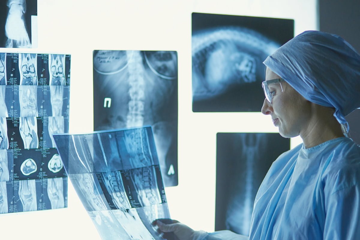
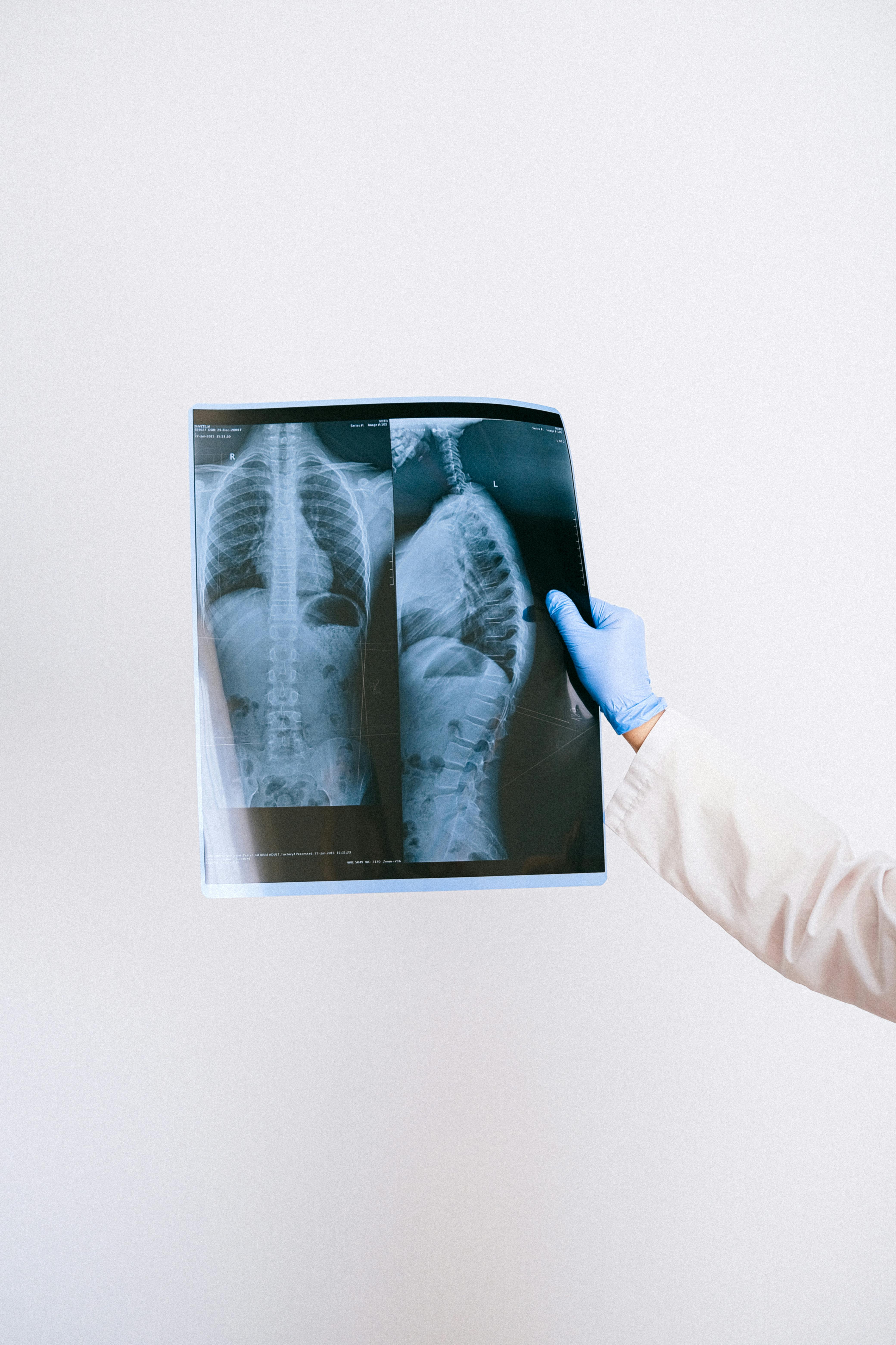
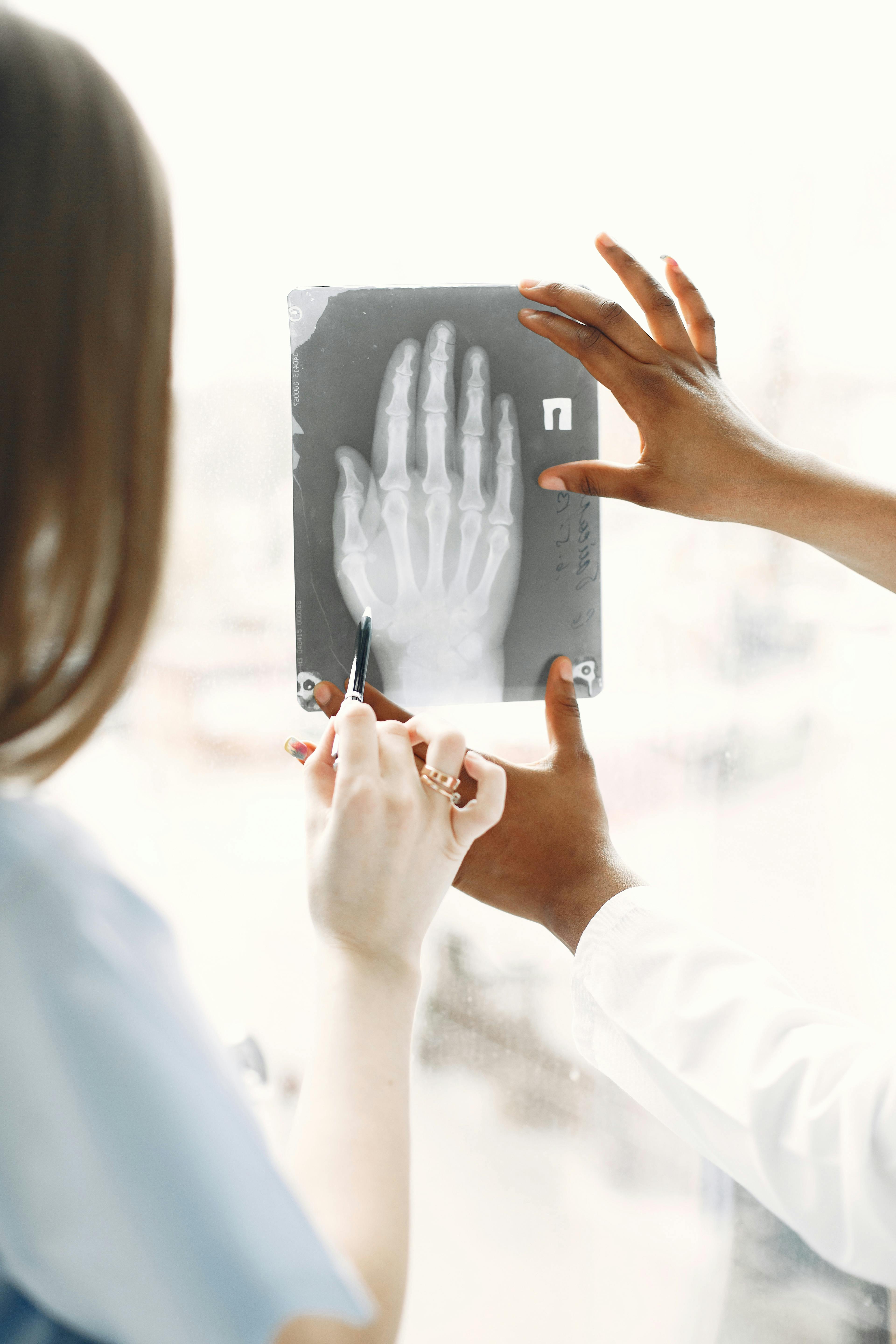
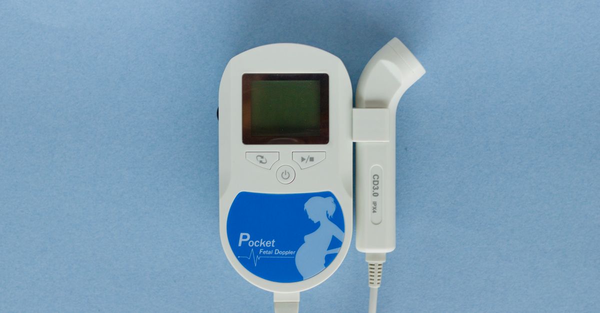
Physician office imaging is evolving rapidly. Traditional setups rely on external radiology labs that incur referral fees, patient travel time, and delayed results. Portable X‑ray units, once limited to hospitals, are now compact, affordable, and easy to deploy in a clinic environment. Similarly, mobile ultrasound devices—handheld or tablet‑based—offer point‑of‑care diagnostics for musculoskeletal injuries, abdominal complaints, and obstetric monitoring.
Despite the clear benefits, many practices hesitate due to perceived costs, regulatory hurdles, and integration challenges. I wanted to quantify how these services could impact workflow, patient satisfaction, and financial performance in a real‑world setting.
Case Study: Implementing Portable Chest X‑Ray Services
Goals and Challenges
The primary objectives were:
- Reduce patient wait times for imaging results.
- Decrease referral costs to external labs.
- Maintain or improve diagnostic accuracy.
Challenges included securing a compliant device, training staff, and ensuring data integration with our electronic health record (EHR).
Implementation Steps
I followed a structured roadmap that can be replicated in other practices:
Step 1: Needs Assessment & Vendor Selection
- Identified imaging frequency and common indications.
- Compared vendors on device size, radiation dose, image quality, and software integration.
- Selected a low‑dose portable X‑ray system with built‑in DICOM export.
Step 2: Regulatory & Safety Compliance
- Obtained necessary state licensing for in‑office radiography.
- Implemented radiation safety protocols, including lead shielding and exposure monitoring.
- Documented SOPs for patient positioning and image acquisition.
Step 3: Workflow Integration
- Allocated a dedicated imaging station near the examination room.
- Updated EHR templates to include imaging orders, results, and billing codes.
- Trained physicians and nurses on device operation and image interpretation basics.
Step 4: Pilot & Feedback Loop
- Ran a 2‑month pilot with 150 patients.
- Collected data on turnaround time, diagnostic concordance, and patient satisfaction.
- Adjusted workflow based on staff feedback and image quality issues.
Mobile Ultrasound Integration
Workflow Adaptation
To complement chest X‑ray services, I introduced a handheld ultrasound unit for common primary care indications. The key was to keep the process seamless:
- Physicians perform scans during the same visit.
- Images are stored directly in the EHR via secure Wi‑Fi transfer.
- Radiology consults remain optional for complex cases.
For example, a patient presenting with right upper quadrant pain can receive an abdominal sweep immediately after history taking. The scan is reviewed on a tablet during the visit, and findings such as gallstones or hepatic cysts are documented in real time, allowing the clinician to prescribe treatment or order labs without a separate imaging appointment.
Staff Training
Effective use of mobile ultrasound requires skill. I organized a two‑day training workshop:
- Basic physics and probe selection.
- Targeted use cases: abdominal pain, shoulder impingement, obstetric dating.
- Hands‑on scanning with supervised practice patients.
To ensure competency, we used simulation phantoms that mimic tissue densities and incorporated a competency checklist. Each operator completed 20 supervised scans before performing independent studies, and periodic refresher sessions were scheduled every six months to maintain proficiency.
Outcomes & Metrics
The results after 12 months were compelling:
- Turnaround time for imaging: Reduced from an average of 5 days to within the same visit.
- Cost savings: Eliminated $8,400 in external lab fees and reduced staff overtime by 15%.
- Diagnostic accuracy: Concordance with external radiology reports exceeded 95%, confirming reliable image quality.
- Patient satisfaction: Survey scores improved from 4.1 to 4.7 out of 5, primarily due to convenience and immediate results.
Financial Analysis & Billing Considerations
Beyond the direct cost savings, integrating on‑site imaging creates new revenue streams that can offset equipment depreciation. Each portable X‑ray scan typically generates a CPT code (e.g., 71017 for chest radiograph) with reimbursement rates averaging $120 in most commercial plans. Over 400 scans per year, this translates to roughly $48,000 in gross billing, of which the practice retains about 70% after payer contracts and equipment amortization.
Similarly, mobile ultrasound examinations can be billed under CPT codes such as 76705 (ultrasound, abdominal) or 76700 (obstetric). Although reimbursement varies, an average of $90 per scan adds a modest yet steady income line. Importantly, the practice avoids the “no‑show” risk associated with external referrals because imaging occurs during the office visit.
Billing accuracy is critical. We instituted quarterly audits to verify that all scans were coded correctly and that modifiers for in‑office radiology services (e.g., modifier 59) were applied consistently, ensuring compliance with payer policies and reducing claim denials.
Quality Assurance & Continuous Improvement
Maintaining high image quality over time requires routine checks. We scheduled monthly phantom studies to verify exposure settings and image sharpness. If a scan failed to meet predefined criteria—such as contrast resolution or field‑of‑view—we retrained the operator and updated SOPs.
We also implemented a feedback loop where radiologists from our tele‑consultation partner reviewed a random sample of scans each quarter. Their reports highlighted subtle artifacts that could be corrected, fostering an environment of continuous learning for the clinical staff.
Common Pitfalls and Mitigation Strategies
Even successful implementations face obstacles. Here are the most frequent issues I observed and how to address them:
- Inadequate radiation safety training: Provide refresher courses annually and maintain a log of exposures.
- Data integration hiccups: Work closely with EHR vendors to ensure DICOM compatibility.
- Resistance from staff: Involve clinicians early in the selection process and highlight workflow benefits.
- Device maintenance: Schedule quarterly servicing and keep spare parts on hand.
- IT security gaps: Conduct regular penetration testing of the imaging network, enforce strong password policies, and train staff on phishing awareness to protect patient data.
Future Outlook: AI & Tele‑Radiology Synergy
The next wave involves integrating artificial intelligence algorithms that flag abnormalities in real time, coupled with tele‑radiology consults for complex cases. In my practice, a pilot AI tool identified lung nodules with 92% sensitivity during chest X‑ray scans, allowing us to triage patients more effectively.
While the technology is promising, it requires robust data governance and continuous validation against ground truth. Practices should adopt an incremental approach—starting with basic image capture and gradually adding advanced analytics as confidence grows. Additionally, staying abreast of evolving reimbursement policies for AI‑assisted imaging will help maximize financial returns without compromising compliance.
What challenges have you faced when integrating portable imaging devices into your practice, and how did you overcome them?Frequently Asked Questions
Browse answers to common questions about our services.