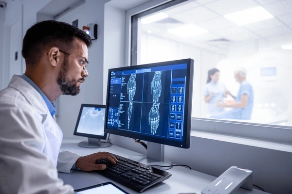
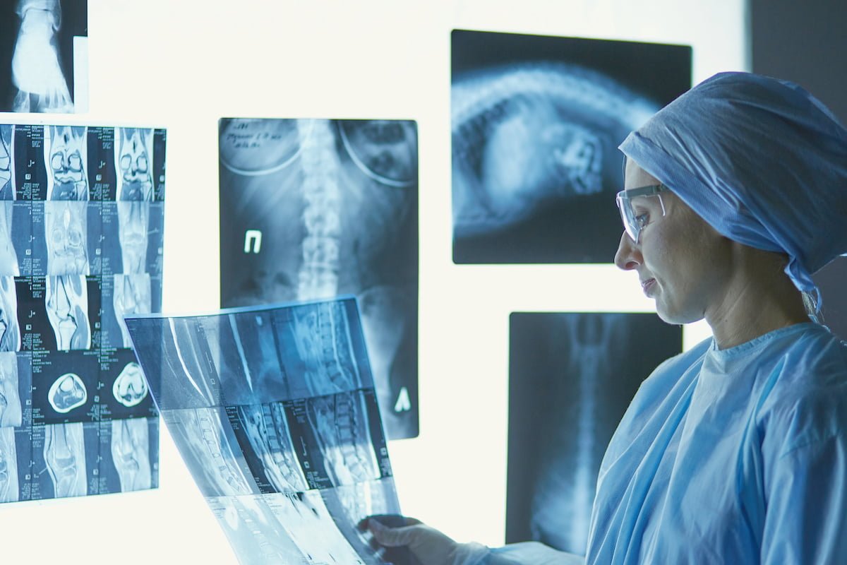
What Is the Role of Regional Mobile Imaging in Modern Healthcare?

In today’s healthcare landscape, patients increasingly expect timely imaging services that come to them rather than requiring travel to distant facilities. Regional mobile imaging—comprising portable ultrasound units, on‑site radiology teams, and mobile X‑ray scanners—addresses this demand by delivering diagnostic quality directly where care is needed. This review examines the core technologies, clinical applications, integration strategies, and economic considerations that define effective regional mobile imaging solutions in 2026.
1. Overview of Regional Mobile Imaging
Regional mobile imaging refers to a coordinated network of imaging services that operate outside traditional hospital or clinic walls. The system typically includes portable ultrasound devices, mobile X‑ray carts, and on‑site radiology personnel who interpret images in real time. By combining these elements, providers can reduce patient transport times, lower readmission rates, and improve diagnostic turnaround.
In 2026, adoption of regional mobile imaging has accelerated due to several factors: advances in wireless data transmission, increased reimbursement for tele‑imaging services, and a growing emphasis on value‑based care. However, success depends on careful attention to equipment selection, staffing models, regulatory compliance, and integration with electronic health records (EHRs).
Key Components of a Regional Mobile Imaging Solution
- High‑resolution portable ultrasound systems with wireless connectivity.
- Mobile X‑ray units that meet radiation safety standards.
- Trained radiology technicians or nurse‑technicians for image acquisition.
- Secure data pipelines linking images to EHRs and PACS.
- Logistics support for vehicle maintenance and equipment transport.
- Reimbursement frameworks aligned with bundled payment models.
Beyond the core hardware, regional mobile imaging relies on a robust operational framework. This includes clear protocols for patient triage, emergency response integration, and data governance to ensure compliance with HIPAA and local privacy regulations. Facilities that invest in staff education around infection control—particularly important during pandemics—can maintain high standards of care while operating outside conventional premises.
2. Portable Ultrasound Imaging: Capabilities and Clinical Impact
Portable ultrasound has evolved from a niche tool to a frontline diagnostic modality in acute care, emergency departments, and remote clinics. Modern devices offer high‑resolution imaging comparable to conventional machines but at a fraction of the cost and footprint. Their battery life and rugged construction enable use in rural hospitals, nursing homes, and even mobile vans.
Despite its strengths, portable ultrasound requires operator skill; image quality can vary with experience. Continuous training programs and remote mentoring can mitigate this risk. Additionally, integration with EHRs enables instant sharing of images for multidisciplinary review.
Clinical Scenarios Where Portable Ultrasound Excels
- Emergency department assessment of abdominal pain (e.g., appendicitis).
- Rapid evaluation of cardiac function in critical care units.
- Guided lumbar punctures and central line insertions.
- Assessment of musculoskeletal injuries on the field or in sports facilities.
- Vascular access planning for dialysis patients.
- Pediatric abdominal examinations to reduce radiation exposure.
In addition, portable ultrasound can support point‑of‑care screening for conditions such as deep vein thrombosis in hospitalized patients, where early detection dramatically changes management. The ability to capture images at the bedside also facilitates interdisciplinary discussions during rounds, fostering a more collaborative approach to patient care.
3. On‑Site Radiology Services & X‑Ray Scanning: How They Work Together
The on‑site radiology model brings a licensed radiologist or a tele‑radiologist’s interpretation directly to the patient’s location. When paired with mobile X‑ray scanners, this approach creates a seamless diagnostic workflow that eliminates waiting times for image review. The combination is especially valuable in long‑term care facilities and rural health centers where specialist availability is limited.
Implementing on‑site radiology requires attention to radiation safety protocols, equipment calibration, and secure data transmission. Compliance with the Radiation Safety Act and local licensing requirements ensures that staff protect both patients and themselves during imaging procedures.
Steps to Launch an On‑Site Radiology Service
- Select a mobile X‑ray unit compliant with current radiation safety standards.
- Hire or contract radiologists capable of remote interpretation via secure video links.
- Integrate the imaging system with the facility’s EHR and PACS.
- Establish clear workflows for image acquisition, transmission, and reporting.
- Conduct pilot testing in a controlled setting before full rollout.
- Create a maintenance schedule to keep equipment calibrated and functional.
Beyond these technical steps, it is essential to develop a quality assurance plan that includes routine audit of image quality, radiation dose monitoring, and feedback loops with interpreting radiologists. This ensures consistent diagnostic accuracy across varied clinical settings.
4. Senior Care Radiology Services: Addressing Elderly Needs
The aging population presents unique challenges for imaging providers. Mobility limitations, polypharmacy risks, and higher prevalence of chronic conditions make it difficult for older adults to travel to imaging centers. Regional mobile imaging solves these barriers by offering on‑site ultrasound and X‑ray services in nursing homes, assisted living communities, and home health settings.
Protocols tailored to seniors emphasize low radiation doses, rapid image acquisition, and minimal patient movement. Portable ultrasound is particularly useful for evaluating hip fractures, soft tissue injuries, or cardiac function without subjecting patients to transport stress.
Best Practices for Senior Care Radiology Services
- Use low‑dose portable X‑ray protocols to minimize radiation exposure.
- Schedule imaging during routine visits to reduce additional appointments.
- Provide staff training on geriatric patient handling and communication.
- Implement a standardized reporting format that highlights findings relevant to elder care clinicians.
- Ensure data security compliance with HIPAA when transmitting images from care facilities.
- Offer post‑imaging follow‑up via telehealth to discuss results with primary physicians.
A practical example is the detection of occult hip fractures in a nursing home after a fall. By performing an ultrasound on site, clinicians can confirm or rule out fracture before arranging transport, thereby reducing the risk of further injury and expediting appropriate orthopedic care.
5. Practical Integration Tips for Clinics and Facilities
Integrating regional mobile imaging into an existing practice requires a structured approach. Begin by assessing current patient flow, IT infrastructure, and staff capacity. Evaluate whether your EHR can interface with portable devices and whether you have the bandwidth to handle increased data traffic.
In my experience, partnering early with vendors that offer end‑to‑end solutions—including equipment, software, and support—reduces onboarding time and minimizes disruptions to patient care. Additionally, establishing clear liability agreements protects both providers and imaging technicians during mobile operations.
Key Considerations When Adding Mobile Imaging
- Compatibility of portable devices with your current PACS/EHR systems.
- Availability of trained personnel for image acquisition and interpretation.
- Insurance coverage and reimbursement policies for mobile services.
- Compliance with state licensing requirements for radiology technicians.
- Infrastructure needs such as Wi‑Fi strength and data security protocols.
- Maintenance contracts and service level agreements (SLAs) with equipment vendors.
A recommended roadmap includes:
- Month 1–2: Conduct a readiness audit covering IT, staffing, and regulatory gaps.
- Month 3–4: Select vendor partners and negotiate SLAs that cover uptime, support response times, and data encryption standards.
- Month 5: Initiate pilot testing in one care setting—such as a regional community hospital—to refine workflows.
- Month 6–7: Expand to additional sites, incorporating lessons learned from the pilot phase.
- Ongoing: Perform quarterly performance reviews focusing on image quality metrics, turnaround times, and patient satisfaction scores.
6. Cost, ROI, and Decision‑Making Factors
The upfront investment for regional mobile imaging includes purchasing portable ultrasound units, mobile X‑ray carts, and securing radiology staff or tele‑radiology contracts. Ongoing costs cover device maintenance, data transmission fees, and training refreshers. However, the potential return on investment (ROI) can be significant if the service reduces readmission rates, shortens ED stays, and attracts new patient populations.
To evaluate ROI, calculate the break‑even point by comparing incremental revenue from new imaging services against total operating costs over a 3‑to‑5‑year horizon. Consider also non‑financial benefits such as improved patient satisfaction scores and reduced transportation costs for patients and families.
Cost Factors to Analyze Before Commitment
- Capital purchase price of portable ultrasound and X‑ray equipment.
- Annual maintenance and software subscription fees.
- Training expenses for technicians and physicians.
- Data storage and transmission bandwidth costs.
- Insurance premiums or liability coverage adjustments.
- Potential reimbursement rates under bundled payment models.
A practical example of ROI calculation might involve a community clinic that adds a portable ultrasound unit. If the unit generates 200 additional studies per year at an average reimbursement of $120 each, the annual revenue is $24,000. Subtracting operating costs ($10,000) yields a net gain of $14,000, implying a payback period of less than two years when combined with savings from avoided patient transports.
Key takeaway: Regional mobile imaging transforms patient access by delivering high‑quality diagnostics directly to care sites, especially benefiting seniors and rural communities while offering a clear path to cost savings when integrated thoughtfully.In conclusion, the convergence of portable ultrasound technology, on‑site radiology teams, and mobile X‑ray scanners provides a flexible, scalable imaging solution that aligns with modern value‑based care goals. By carefully selecting equipment, training staff, ensuring regulatory compliance, and conducting rigorous cost analyses, providers can harness regional mobile imaging to improve clinical outcomes and operational efficiency.
How have you integrated portable imaging into your practice, and what challenges did you face during the transition?Frequently Asked Questions
Browse answers to common questions about our services.